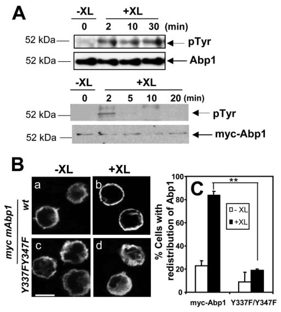
FIGURE 5.
BCR-induced cellular redistribution of Abp1 is dependent on BCR-induced tyrosine phosphorylation of Abp1. A, Untransfected A20 B cells (top) and A20 B cells transfected with myc-Abp1 (bottom) were treated (+XL) or untreated (−XL) with goat anti-mouse IgG for varying lengths of time to activate the BCR. Then cells were lysed, and endogenous Abp1 and myc-Abp1 were purified from cell lysates by immunoprecipitation using anti-Abp1 and anti-myc Abs, respectively. The immunoprecipitates were analyzed by SDS-PAGE and Western blot, probing with anti-phosphotyrosine mAb (4G10). The blots were stripped and reblotted with anti-Abp1 or anti-myc Ab. Shown are representative blots of three independent experiments. B, A20 cells transiently transfected with wt myc-Abp1 (Ba-Bb) and myc-Abp1 Y337F/Y347F (Bc-Bd) were treated (+XL) and untreated (−XL) with goat anti-mouse IgG for 2 min, and then fixed, permeabilized, and labeled with Cy3 anti-myc mAb for myc-Abp1. Cells were analyzed using a confocal fluorescence microscope. Shown are representative images of three independent experiments. Bar, 10 µm. C, Cells in images were quantified, as described in Fig. 4B. Over 100 cells from three independent experiments were analyzed. Shown are the averages (±SE) from three independent experiments. **, p < 0.01.

