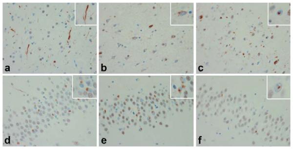
Figure 2.
Cortex (a, b & c) and dentate gyrus (d, e & f) of Type 1 (a & d), Type 2 (b & e), and Type 3 (c & f) FTLD-U according to Sampathu & Cairns [1]. Type 1. Predominance of dystrophic neurites (DN) (a, inset) in cortex with round neuronal cytoplasmic inclusions (NCI) (d, inset) in the dentate gyrus. Type 2. Predominance of NCI, including granular cytoplasmic TDP-43 immunoreactivity, with sparse DN in cortex (b, inset) and dentate gyrus (e, inset). Type 3. NCI, DN & neuronal intranuclear inclusions (NII) (c, inset) and NCI and NII in dentate gyrus (f, inset).

