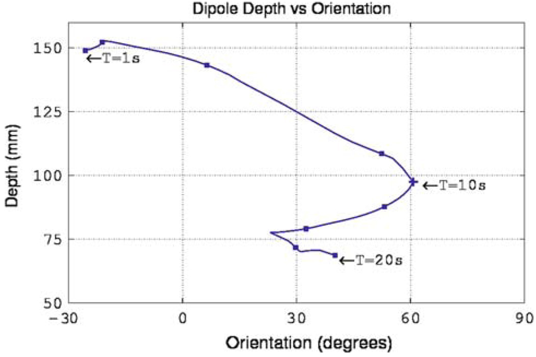
FIGURE 7.
Variation of depth and orientation of the single realistic dipole source (as presented in Fig. 1 and Fig. 6) over one slow wave cycle. The source initiates at the mid-corpus region of the stomach (located furthest from the sensor) and progresses toward the antrum (located closest to the sensor) while the dipole orientation oscillates between −30° and 60° through the cycle.

