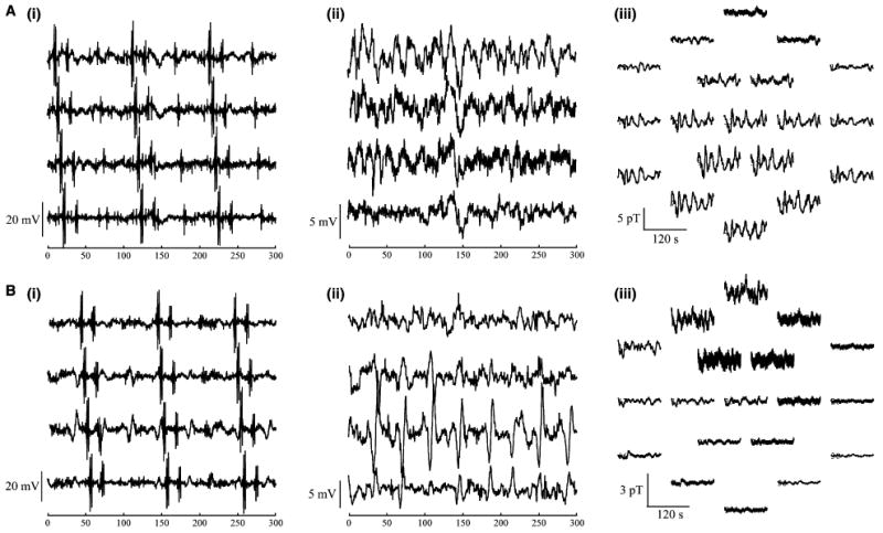
Figure 2.

Recordings (A) before and (B) after gastric division in (i) raw and (ii) filtered serosal electrodes and in (i) the multichannel SQUID magnetometer. Recordings from four sequential electrodes demonstrate propagation of spiking activity in unfiltered data (i). In panel (Aii) and (Bii) the spiking activity has been filtered to show only slow waves. Magnetogastrogram signals are mapped to the spatial location of the sensors (Aiii) before and (Biii) after gastric division.
