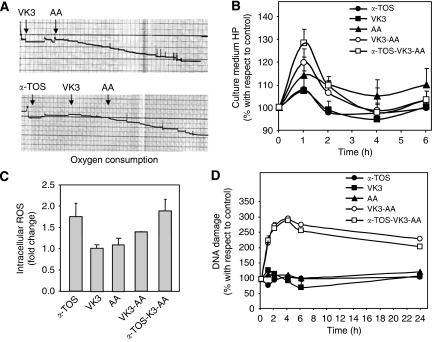
Figure 5.
Reactive oxygen species (ROS) generation and DNA fragmentation induced in prostate cancer cells by α-tocopheryl succinate (α-TOS) or vitamin K3 (VK3) plus ascorbic acid (AA) alone or in combination. (A) The capacity of α-TOS or VK3 plus AA alone or in combination to induce ROS formation was evaluated by assessing the oxygen consumption using the Clark's oxygen electrode. (B) PC3 cells were placed in 96-well flat-bottom tissue culture plate at 104 per well. After overnight incubation, cells were treated with α-TOS (30 μM), VK3 (3 μM), and AA (0.4 mM) alone or in combination, and aliquots of the conditions media were taken at regular intervals and evaluated for the level of hydroperoxides using the d-ROMs assay. Data are expressed as of percentage variation with respect to the control. (C) Intracellular ROS were estimated using the fluorescent dye 2′7′-dichlorofluorescein diacetate (DCFA). PC3 cells were seeded in 24-well flat-bottom plates and 20 μM of DCFA added. After 30 min of incubation, the cells were exposed to α-TOS (30 μM), VK3 (3 μM), and AA (0.4 mM) alone or in combination. After 24 h, the cells were evaluated by flow cytometry. The amount of ROS was detected as fluorescence intensity normalised for the blank (samples without DCFA) and expressed as fold change with respect to the control (untreated cells). (D) DNA fragmentation was analysed using the alkaline comet assay. PC3 cells were seeded in 96-well flat-bottom tissue culture plates at 104 per well, treated after overnight incubation with α-TOS (30 μM), VK3 (3 μM), and AA (0.4 mM) alone or in combination, and evaluated for DNA damage at different time points. The level of DNA strand breaks was assessed using the comet assay that is based on visual scoring with the maximum of 400 arbitrary units (see Material and Methods for detail). The data are expressed as the percentage variation with respect to the control (untreated cells).

