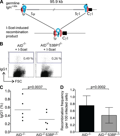
Figure 2.
Loss of 53BP1 decreases the joining efficiency of two distal I-SceI–induced DSBs. (A) Schematic representation of the IgHI-96k allele (top) and the I-SceI–induced recombinant that encodes IgG1 (bottom). LoxP sites are indicated as red triangles, and I-SceI sites are indicated as blue circles. (B) Representative flow cytometry experiments showing CSR to IgG1 of IgHI-96kAID−/− and IgHI-96kAID−/−53BP1−/− B cells 72 h after the first infection with an I-SceI–encoding retrovirus. (C) Graph shows the results of five independent flow cytometry experiments, with each dot representing an individual experiment. The p-value was calculated using a two-tailed paired Student’s t test. The means are shown as horizontal lines. (D) Bar graph showing I-SceI to I-SceI recombination frequency in the presence and absence of 53BP1, determined by nine independent PCR experiments. Error bars indicate standard deviation. The p-value was calculated using a two-tailed paired Student’s t test. FSC, forward scatter.

