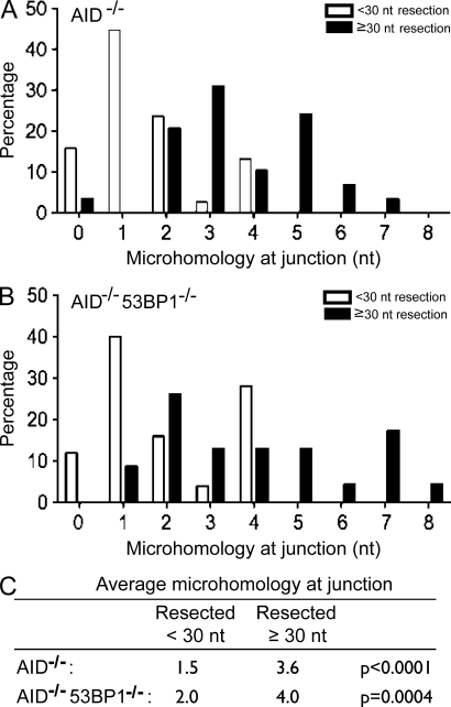
Figure 4.
Resected DSBs are joined using microhomology-mediated end joining independently of 53BP1. (A) Bar graph shows microhomology at junctions of PCR products cloned from I-SceI–infected IgHI-96kAID−/− B cells. White bars indicate sequences with little resection (<30 nt), and black bars indicate sequences with resection ≥30 nt. A total of 67 individual sequences were analyzed from three different mice. (B) Bar graph shows microhomology at junctions of PCR products cloned from I-SceI–infected IgHI-96kAID−/−53BP1−/− B cells. White bars indicate sequences with little resection (<30 nt), and black bars indicate sequences with resection ≥30 nt. A total of 48 individual sequences were analyzed from three different mice. (C) Table indicates the mean number of nucleotides of microhomology at junctions in I-SceI–infected IgHI-96kAID−/− and IgHI-96kAID−/−53BP1−/− B cells.

