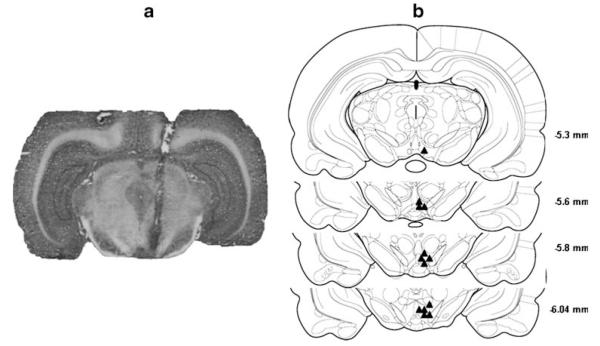
Fig. 1.
a A photomicrograph of a representative brain slice with an injection site within the p-VTA. b Representative non-overlapping placements of the injection sites within the p-VTA. The p-VTA corresponds to coronal sections from 5.3 to 6.0 mm posterior to bregma (Rodd-Henricks et al. 2000). The filled triangles represent the injection sites

