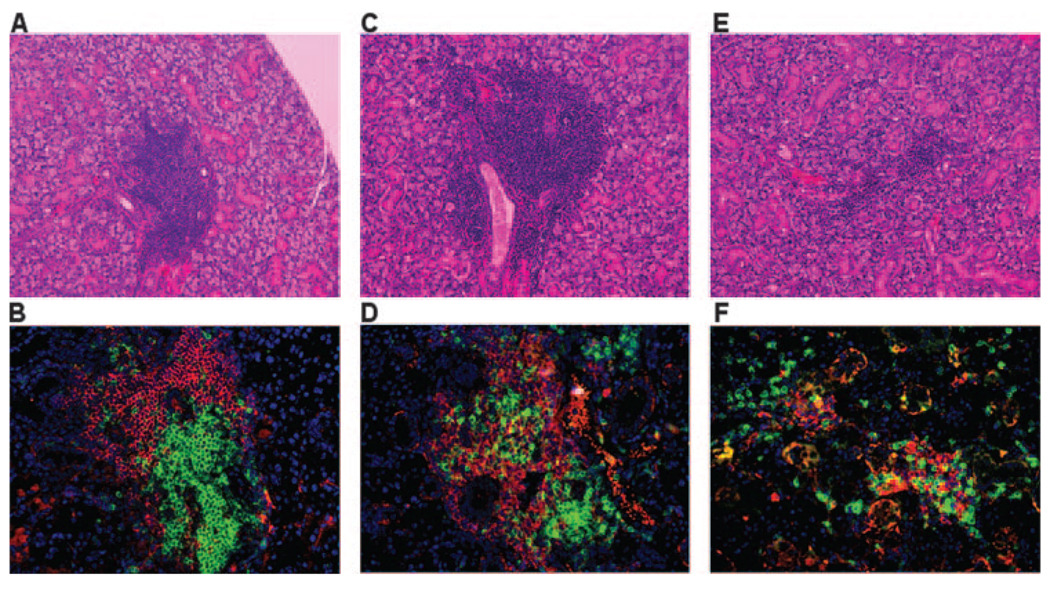
FIGURE 3.
Histological examination of the exocrine glands. Submandibular glands removed from groups of mice at ages ranging from 20 to 28 wk were fixed in 10% formalin. Each gland was serially sectioned (5-µm thickness) and histology performed on two sections cut 50 µm apart. Each section was stained with Mayer’s H&E dye (A, C, and E) or immunofluorescent Abs for determining the distribution of B (anti-B220, red) and T (anti-CD3, green) cells in lymphocytic foci (B, D, and F). The sections were counterstained with 4′,6′-diamidino-2-phenylindole (DAPI) (blue). The numbers of focus were counted across whole histological sections of glands from 20-wk-old female NOD.B10-H2b mice (n = 8; A and B), 28-wk-old female NOD.B10-H2b.C-Stat6+/+ mice (n = 18; C and D), and 25-wk-old female NOD.B10-H2b.C-Stat6−/− mice (n = 23; E and F). All H&E sections and immunofluorescent staining was examined at ×200 magnification. The number of foci for the two sections was averaged for comparisons.

