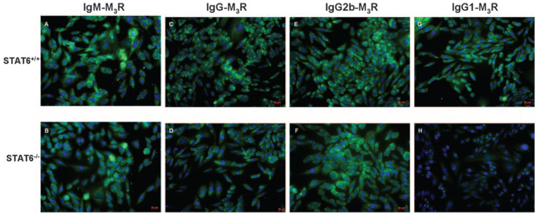
FIGURE 6.
Detection of M3R isotypic autoantibodies by immunofluorescence. Sera collected from NOD.B10-H2b.C-Stat6+/+ and NOD.B10-H2b.C-Stat6−/− mice were incubated at a dilution of 1/50 with newly cloned M3R-transfected Flp-In CHO cells (see Ref. 14 and Ref. 19) for 1 h in a humidified chamber at room temperature. The cells were washed five times (5 min each wash) with PBS, then incubated 30 min at room temperature with FITC-conjugated secondary Abs specific for IgM (A and B), IgG (C and D), IgG2b (E and F), or IgG1 (G and H) isotypes diluted 1/100 (Serotec). Cells were again washed and visualized using a Zeiss Axiovert 200 M microscope at ×100 magnification. Images represent an exposure time of 25 ms.

