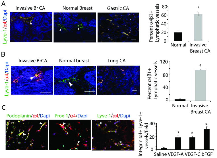
Figure 1. Integrin α4β1 and fibronectin are markers of proliferative lymphatic endothelium in human and murine tumors.
(A) Left, integrin α4β1 (red), Lyve1 (green) and DAPI (blue) immunostaining of human invasive breast ductal carcinoma, normal breast and gastric tumors. Right, percent integrin α4+ lymphatic vessels +/- SEM per 100× field in mammary tissue from A (n=15 normal, 35 invasive, *p<0.001). (B) Left, integrin α4β1 (red), Lyve1 (green) and DAPI (blue) immunostaining of mammary glands from PyMT- (normal) and PyMT+ (invasive breast carcinoma) mice, and LLC tumors. Right, percent +/- SEM integrin α4+ lymphatic vessels per 100× microscopic field in PyMT- and PyMT+ mammary tissue (n=10, *p<0.001). (C) Left, immunostaining of VEGF-C treated tissue for Lyve-1, podoplanin or Prox-1 (green), integrin α4β1 (red), and DAPI (blue). Right, mean number of integrin α4+ lymphatic vessels/100× microscopic field in saline, bFGF, VEGF-A or VEGF-C saturated Matrigel (n=10, *p<0.001). Scale bars, 50μm.

