
Figure 5.
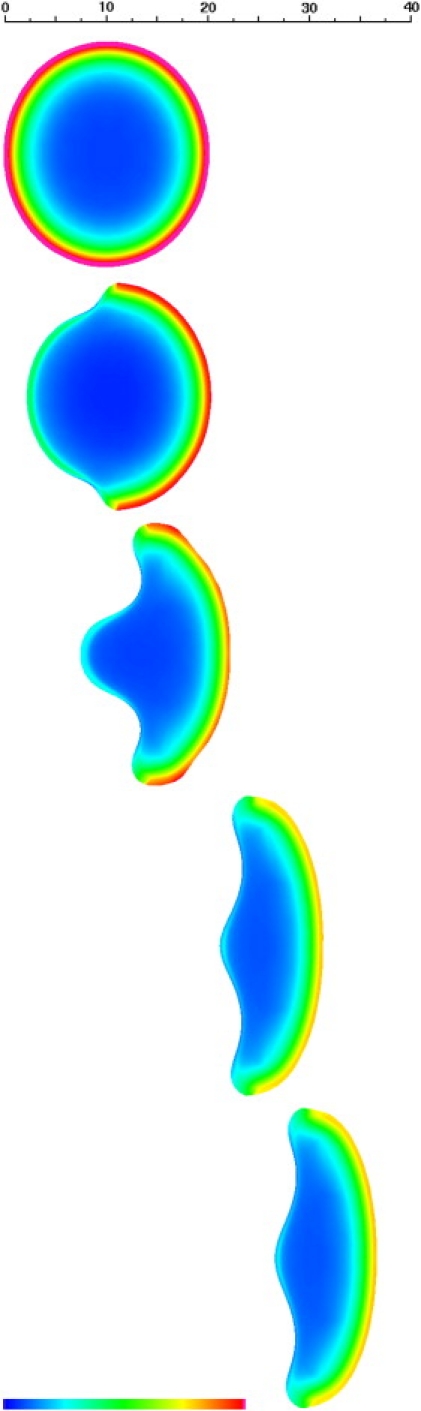
Ventral cytoskeletal volume fraction for the keratocyte at 0, 10, 50, 150, and 200 s. Distance scale in μm, color scale spans 0–2% volume fraction. Note the decrease in maximum cytoskeletal density between the spread discoid cell and the migrating keratocyte. This is due to the fact that in the keratocyte, the activated leading-edge moves sufficiently fast as to prevent full polymerization of cytoskeleton at a given location. See Movie S4.