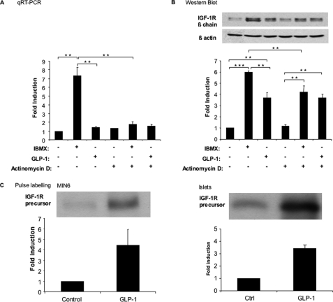
FIGURE 4.
IBMX and GLP-1 regulate IGF-1R expression by transcriptional and post-transcriptional mechanisms. A and B, MIN6 cells were exposed to either IBMX (10 μm) or GLP-1 (100 nm) for 18 h and in the presence or absence of actinomycin D (1 μg/ml). IGF1-R mRNA (A) as well as protein (B) expression was quantitated by qRT-PCR and Western blot analysis, respectively. C, MIN6 cells (left panel) or mouse islets (right panel) were biosynthetically labeled for 1 h with [35S]methionine and [35S]cysteine in the presence or absence of GLP-1 (100 nm). IGF-1R was immunoprecipitated from the cell lysates and separated by gel electrophoresis; the amount of newly synthesized precursor receptor was quantitated by densitometric analysis of the autoradiograms. The data are the means ± S.D. from three independent experiments. **, p < 0.01; ***, p < 0.001. Ctrl, control.

