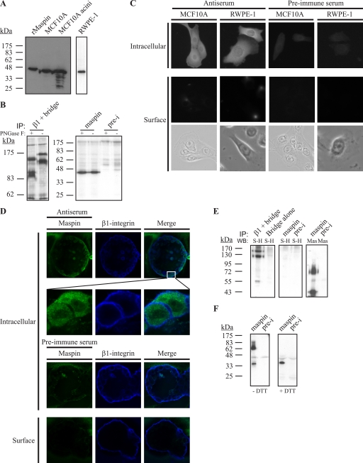
FIGURE 1.
Maspin is expressed by MCF10A and RWPE-1 cells but is absent from the cell surface. A, lysates of MCF10A cells and acini or RWPE-1 cells were separated via 12.5% SDS-PAGE and immunoblotted with recombinant maspin (rMaspin) as a control. The membrane was probed with mouse anti-maspin monoclonal antibody diluted 1:1000 and detected with horseradish peroxidase-conjugated secondary against mouse IgG. RWPE-1 lysates were analyzed on a separate gel. B, maspin in MCF10A cells is not glycosylated. Lysates of 30-min metabolically labeled MCF10A cells were immunoprecipitated (IP) using mouse anti-β1-integrin monoclonal antibody (left panel), rabbit anti-maspin polyclonal antiserum (right panel), or preimmune (pre-i) serum and then either treated with PNGase F or left untreated. Maspin samples were separated by 12.5% SDS-PAGE, and β1-integrin samples were separated by 7.5% SDS-PAGE. Gels were analyzed by fluorography. C, maspin is in the nucleus and cytoplasm and not on the cell surface. MCF10A and RWPE-1 cells were fixed and permeabilized or fixed alone to detect cell surface proteins and then probed with mouse anti-maspin polyclonal antiserum or with the preimmune serum. The primary antibody was detected using goat Alexa 488-conjugated secondary antibodies. Cells were examined by epifluorescence microscopy. Brightfield images of cells examined for cell surface staining are shown. D, maspin is intracellular in each luminal epithelial cell and does not co-localize with β1-integrin. MCF10A cells were grown on Matrigel, and acini were developed for 20 days. Acini were prepared as described for B and examined by confocal microscopy. Images shown are single optical sections. E, MCF10A cells were surface-biotinylated, and lysates were immunoprecipitated using rabbit anti-maspin polyclonal antiserum, preimmune serum, or mouse anti-β1-integrin monoclonal antibody. Immune complexes were collected and analyzed by 10% SDS-PAGE, and the immunoblot (WB) was probed with horseradish peroxidase-conjugated streptavidin (S-H) diluted 1:5000. The blot was stripped and re-probed with mouse anti-maspin monoclonal antibody (Mas) and detected with horseradish peroxidase-conjugated secondary against mouse IgG. Bridge refers to samples immunoprecipitated with bridging antibody alone. F, MCF10A cell lysates were immunoprecipitated, and immune complexes were left nonreduced (without dithiothreitol (−DTT)) or reduced (+DTT) and analyzed via 12.5% SDS-PAGE and immunoblotting with mouse anti-maspin monoclonal antibody followed by horseradish peroxidase-conjugated secondary against mouse IgG.

