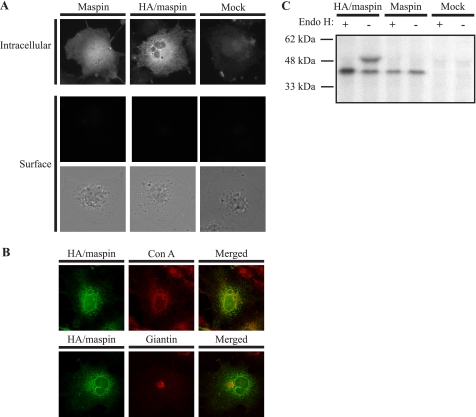
FIGURE 4.
Glycosylated maspin is retained in the ER. A, COS-1 cells transfected with HA/maspin or maspin were prepared as described in the legend to Fig. 1 and probed with mouse anti-maspin polyclonal antiserum. As a control, COS-1 cells were mock-transfected. The primary antibody was detected with goat anti-mouse IgG conjugated to Alexa 488, and cells were examined by epifluorescence microscopy. The lower panel shows brightfield images of cells examined for cell surface staining. B, COS-1 cells transfected with HA/maspin were fixed, permeabilized, and probed with mouse anti-maspin monoclonal antibody. The primary antibody was detected with goat anti-mouse IgG conjugated to Alexa 488. The ER was marked with Alexa Fluor 594-conjugated concanavalin A (Con A), and the Golgi was marked with rabbit anti-giantin polyclonal antibody detected with goat anti-rabbit IgG conjugated to RITC. Cells were examined by confocal microscopy. Images show single optical sections. C, COS-1 cells were transfected with HA/maspin or maspin DNA or without DNA (Mock). Forty-eight hours after transfection, cells were labeled for 30 min in medium lacking methionine containing 100 μCi of [35S]methionine and then incubated in complete medium for 6 h. Cell lysates and medium were then collected and immunoprecipitated with rabbit anti-maspin polyclonal antiserum. Immune complexes were either treated (+) or left untreated (−) with Endo H. Samples were then reduced and analyzed by 10% SDS-PAGE and fluorography.

