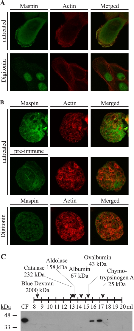
FIGURE 5.
Maspin is not stably associated with the cytoskeleton or other cellular proteins and exists as a monomeric serpin. A and B, maspin is a soluble cytoplasmic protein. MCF10A monolayers (A) growing on microscope slides and MCF10A acini (B) were extracted with HMKE buffer containing digitonin and then fixed, permeabilized, and probed with mouse anti-maspin polyclonal antiserum. The primary antibody was detected with goat anti-mouse IgG conjugated to Alexa 488. The actin cytoskeleton was marked by RITC-conjugated phalloidin. Cells were examined by confocal microscopy. Images show single optical sections. C, MCF10A cells were harvested in hypotonic buffer, and gel filtration was performed on the cytosolic fraction (CF) of lysate. Fractions were separated by 12.5% SDS-PAGE, analyzed by immunoblotting for maspin using mouse anti-maspin monoclonal antibody, and detected with horseradish peroxidase-conjugated secondary against mouse IgG. A series of molecular mass standards ranging between 25 and 2000 kDa was assessed, and the fractions in which they appeared are shown. The molecular mass of the eluted maspin was within the predicted monomeric range.

