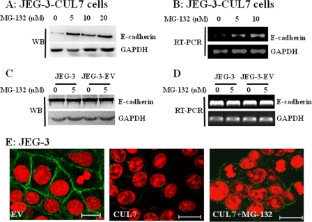
FIGURE 3.
MG-132 partially restored E-cadherin expression in JEG-3 cells stably transfected with CUL7. JEG-3 cells stably expressing CUL7 (A and B), parental JEG-3 cells, and JEG-3 cells stably transfected with empty vector (C and D) were treated with the indicated concentrations of MG-132 for 6 h (RT-PCR) and 24 h (WB) and subjected to RT-PCR (B and D) and Western blotting (A and C) analysis of E-cadherin expression, respectively. E is an immunofluorescence micrograph showing the expression of E-cadherin (green) in JEG-3 cells transfected with EV (left panel), JEG-3 cells transfected with CUL7 (middle panel), or JEG-3-CUL7 cells treated with 10 μm MG-132 for 24 h. Nuclei were shown by PI staining. Bar = 20 μm.

