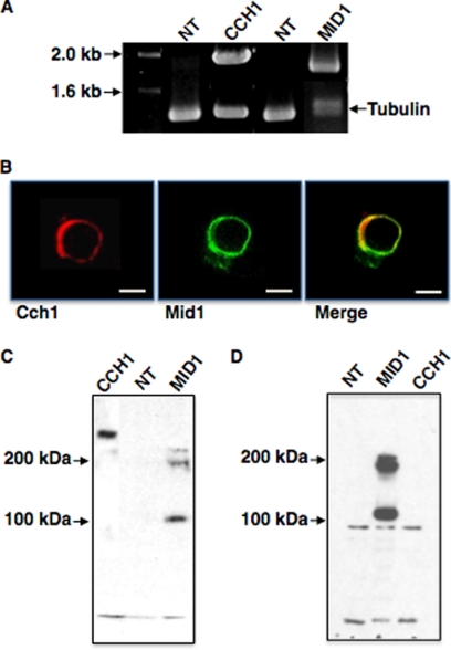
FIGURE 1.
Cch1 and Mid1 proteins are expressed in the plasma membrane of HEK293 cells. A, reverse transcription-PCR analysis detected transcripts of CCH1 and MID1 in HEK293 cells previously transfected with CCH1 and MID1 cDNA. Transcripts were not detected in nontransfected (NT) cells. Tubulin is shown as a loading control. B, Mid1 was tagged with a C-terminal GFP tag and visualized by confocal microscopy (center panel). Mid1 is expressed predominantly on the cell surface of HEK293 cells, and Mid1 co-localizes with Cch1 (right panel, merge). Immunofluorescence (Texas Red) of Cch1 (nontagged) revealed a surface distribution in HEK cells (left panel) similar to Mid1. A primary peptide antibody to the C terminus of Cch1 and a Texas Red-conjugated secondary antibody were used to visualize Cch1. Scale bars, 10 μm. C, Western blot analysis of isolated plasma membranes from HEK293 cells expressing Cch1 or Mid1 and revealed polypeptides corresponding to the predicted sizes for Cch1 (∼220 kDa) and Mid1 (monomer, ∼100 kDa; complex, ∼200 kDa). D, Western blot analysis of surface biotinylation of HEK293 cells expressing Cch1 or Mid1 revealed two distinct bands unique to the lane corresponding to Mid1 (monomer, ∼100 kDa; complex, ∼200 kDa). These bands were not detected in nontransfected HEK cells (NT). A protein band corresponding to biotinylated Cch1 could not be detected.

