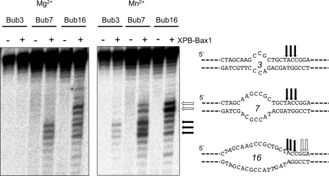
FIGURE 2.
XPB-Bax1 cleaves a range of bubble substrates. XPB-Bax1 cleaves bubbles of 7 and 16 nt in the presence of magnesium and ATP and additionally a bubble of 3 nt in the presence of manganese and ATP. The cleavage sites are shown mapped for the three substrates. The black arrows show cleavage sites in common for all three bubbles. Cleavage further into the duplex 3′ of the bubble (white arrows) suggests further opening of the DNA by XBP.

