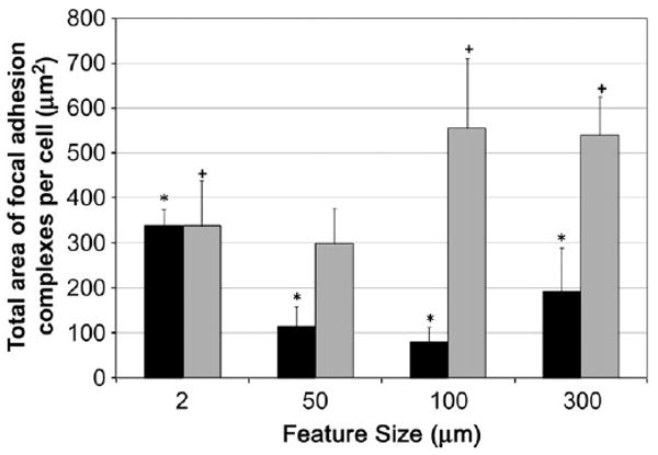
Fig. 8.

Focal adhesion complex (FAC) characterization of adherent cells on NP-modified surfaces and unmodified glass. FAC data for BAEC (black bars) and MC3T3 (gray bars) cells as cultured on glass, 50, 100 and 300 nm silica NP-modified glass surfaces. Total area of focal adhesion complexes per cell are reported as an average from 10 cells per NP substrate for each cell type. The feature size on glass surface is 2 nm. The asterisks (*) indicate statistical significance (p < 0.05) for all BAEC cultured on NP-modified surfaces when compared to the glass control. A plus (+) symbol indicates statistical significance (p < 0.05) for MC3T3 cultured on NP-modified surfaces when compared to the glass control.
