Fig. 1.

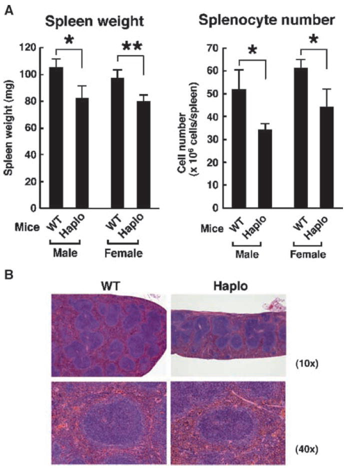
Characteristics of spleens from brx+/− mice. (A) The spleens from brx+/− (Haplo) mice weighed less and contained fewer splenocytes compared with those of WT mice. Data in the left panel represent the mean spleen weight, with error bars indicating the SEM (Haplo mice: male, n = 5; female, n = 7; WT mice: male, n = 5; female, n = 7), whereas those in the right panel represent the mean ± SEM of splenic mononuclear cells (Haplo mice: male, n = 5; female, n = 7; WT mice: male, n = 5; female, n = 7). *P < 0.05; **P < 0.01. (B) brx+/− mice exhibited an altered splenic follicular structure compared with that of WT mice. Results of hematoxylin and eosin (H&E) staining are shown and are representative of three experiments. The spleens of brx+/− mice had smaller follicles than did those of WT mice. Top and bottom panels show low- (10×) and high-magnification (40×) images, respectively.
