
Figure 1.
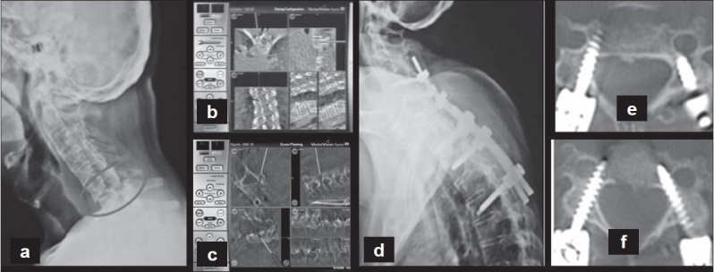
(a) Preoperative lateral radiograph of cervical spine showing a C6-7 chance fracture in a patient with ankylosing spondylitis (b, c) Intra operative navigation images of thoracic and cervical spine- pedicle entry point and trajectory and planning of screws (d, e, f) Postoperative radiograph and CT scan showing the placement of the screws