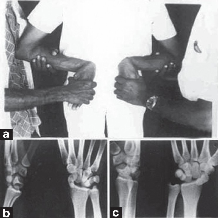
Figure 1.

(a) Clinical photograph showing the position of limbs held on back in forcible full volar flexion, radial deviation and pronation at the wrists.(b) Lateral and anteroposterior X-rays of left and right.(c) wrists showing posterior perilunate dislocation. Note: Irregularity of posterior horn of lunate in lateral view
