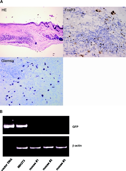
Figure 5.
Donor skin grafts of Treg-treated chimeras show high frequencies of mast cells and Tregs. (A) Histopathology of donor skin grafts from Treg-treated chimeras revealed high frequencies of mast cells (Giemsa) and FoxP3 positive cells (immunohistochemistry) (HE, hematoxylin and eosin stain, magnification 100×; FoxP3, immunohistochemistry with specific FoxP3 antibody, magnification 400×; Giemsa, Giemsa staining, magnification 200×; representative graft shown 5 months post-BMT). (B) Genomic DNA isolated from skin grafts of FoxP3-Treg treated mice (n = 3) was subjected to PCR analysis specific for GFP. Grafts lacked detectable GFP expression, indicating that graft-infiltrating FoxP3 Tregs did not originate from the therapeutically administered Tregs. FoxP3-transduced NIH3T3 cells and FoxP3 vector were used as positive controls. DNA levels of GFP (upper panel) and β-actin (lower panel) are shown.

