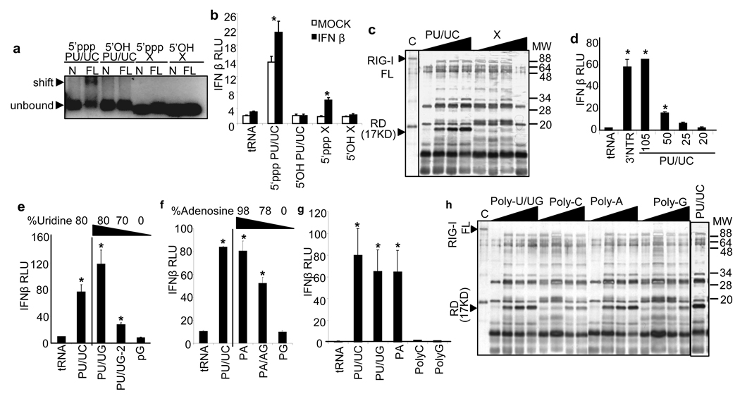
Figure 3. Poly-uridine and poly-adenosine ribonucleotides are RIG-I ligands.
a, Gel shift analysis of complex formation between 25 pmol of purified N-RIG (control) or full-length RIG-I (FL) and 10 pmol of poly-U/UC (PU/UC) or X region RNA containing 5’ppp or 5’OH as indicated. Arrows denote position of unbound RNA and RNA/RIG-I complexes. b, Effect of 5’ppp on IFN-β promoter activity. Huh7 cells were either mock-treated or treated with IFN-β 8 h prior to transfection with 1 µg (30 pmol ) of RNA. c, Effect of poly-U/UC or X region RNA on RIG-I activation. The silver-stained gel image shows trypsin-digestion products of RIG-I that was pre-incubated with increasing amounts poly-U/UC or X region RNA. Arrows indicate positions of full length (FL) RIG-I and the 17 kDa trypsin-resistant RD of from RIG-I/RNA complexes. d, Effect of nt length of 1 µg (30-150 pmol) poly-U/UC 3’ truncation products on IFN-β promoter signaling in Huh7 cells. e–g, Effect of nt composition on IFN-β promoter signaling in Huh7 cells transfected with 1 µg (30 pmol ) of RNA. h, Effect of nt composition on RIG-I activation. The silver-stained gel image shows trypsin-digestion products of RIG-I that was pre-incubated with increasing amounts poly-U/UG, poly-C, poly-A, or poly-G RNA. Arrows indicate positions of full length RIG-I and the 17 kDa trypsin-resistant RD. We confirmed the 17 kDa fragment as the RIG-I RD by immunoblot analysis of the digestion products using an antiserum specific to the RIG-I carboxyl terminus (not shown), as previously described 17. Asterisks indicate significant difference (P<0.01) as determined by Student’s T-test.

