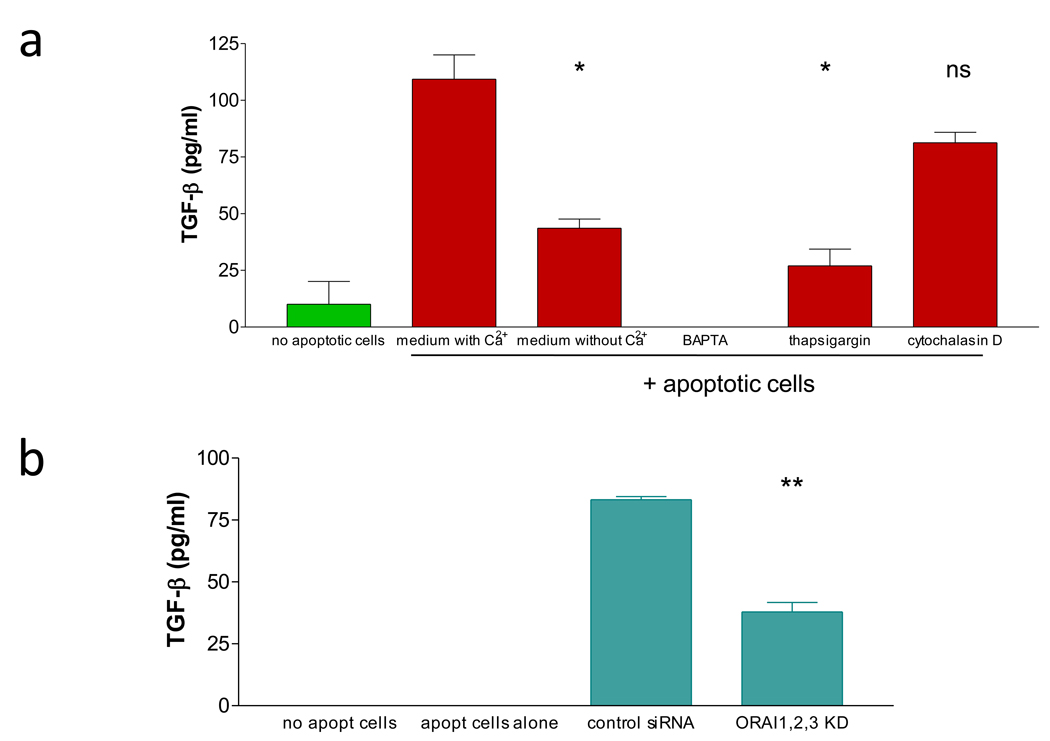
Figure 5. Both intracellular and extracellular Ca2+ is crucial for TGF-β secretion by phagocytes encountering apoptotic cells.
a) J774 phagocytes cells were incubated with apoptotic thymocytes in the presence of absence of calcium in the medium, or in the presence of calcium but with calcium inhibitor drugs. The supernatants were collected and analyzed for TGF-β levels by ELISA. Drugs were added at the same time as the apoptotic cells.
b) NIH3T3 cells transfected with control or orai1, orai2 and orai3 specific siRNA were incubated with apoptotic thymocytes and secretion of TGF-β was measured in the medium 24 hrs later. No apopt cells shows the amount of TGF-β secreted by NIH3T3 cells without addition of apoptotic thymocytes, while apopt cells alone is the amount of TGF-β in wells with apoptotic thymocytes without any phagocytes.
Each figure is representative of at least three independent experiments. Significance is indicated as follows: p>0.05 – (ns), 0.01–0.05 – (*), 0.001–0.01 – (**), <0.001 – (***).

