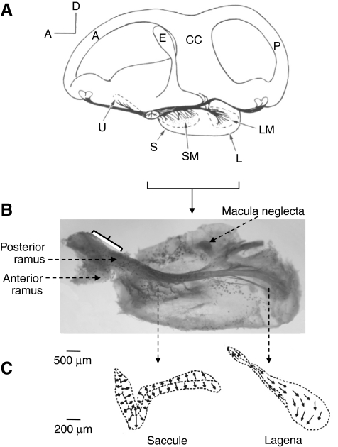
Fig. 1.
(A) Diagram of a medial view of the right ear showing the spatial orientation of the saccule and lagena (located vertically in one pouch) in Acipenser sturio (Baltic sturgeon) (Retzius, 1881). A, anterior semicircular canal; CC, crus commune; E, endolymphatic duct; L, lagena; LM, lagenar macula (epithelium); P, posterior semicircular canal; S, saccule; SM, saccular macula; U, utricle. (B) Innervation of the right saccule and lagena in Acipenser fulvescens (medial view; photograph by M. Meyer). The bracket indicates the recording site on the posterior ramus carrying fibers from the saccule and lagena. The more posterior portion of the nerve (just innervating the lagena) is tightly attached to the pouch making recordings difficult without damaging the pouch. (C) Outline of the saccule and lagena in A. fulvescens showing the orientation of the hair cells. Arrows are pointing in the direction of the kinocilium [adapted from Lovell et al. (Lovell et al., 2005)].

