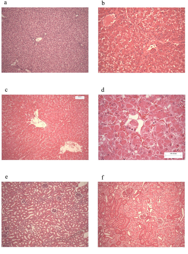
Figure 9.
Histology. This figure depicts representative photographs of liver and renal sections. Panel a, represents liver tissue taken from a control pig. Panel b represents liver tissue taken from pig 4 and demonstrates moderate injury. There is diffuse microvesicular change, with moderately severe centrilobular necrosis. Panels c and d (higher power) represents liver tissue taken from pig 7 demonstrating more severe coagulative centrilobular necrosis. Panel e represents renal tissue taken from a control pig. Panel f represents liver tissue taken from pig 7 demonstrating severe vacuolar injury to the cortical tubules in keeping with the development of acute tubular necrosis.

