
Figure 6.
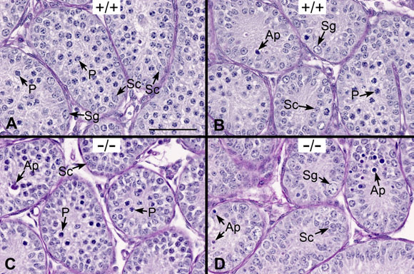
Post-natal day 10 tubule cross-sections of Ehd1-/- male mouse testes show no major lesions. Day 10 testes were Bouin's-fixed, PAS-stained and hematoxylin-counter-stained to visualize the glycoproteins/acrosomes (pink) and nuclei (blue) and analyzed by light microscopy using a 40× objective lens. Stages are labeled with Roman numerals. (A, B) The seminiferous tubules of WT (+/+) mice exhibit Sertoli cell nuclei (Sc) near the basement membrane or toward the lumen, large spermatogonia (Sg) near the basement membrane, pachytene spermatocytes (P) and occasional apoptotic-like (Ap) nuclei near the lumen. (C, D) The seminiferous tubules of Ehd1-/- (-/-) mice are shown for comparison. Scale bar in A = 50 μm for A-D.