Figure 1.

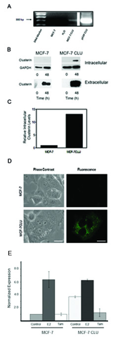
Over-expression of clusterin in MCF-7 cells. Panel A: Stable transfection of clusterin into MCF-7 cells using the Gateway pDEST-clusterin, verified by PCR amplification. Following stable transfection into MCF-7 cells using the pDEST-CLU generated by the Gateway technology, nuclear DNA was prepared from MCF-7 and MCF-7CLU cells and analyzed for plasmid integration by PCR amplification using clusterin and vector DNA sequences derived from the clusterin cDNA and the vector. The pEXP-clusterin was used as a positive control. Panel B: Cell lysates (30 micrograms) and conditioned medium (50 microliters) prepared from MCF-7 and MCF-7CLU before and after 48 h of incubation in serum free medium were separated on 12.5% SDS-PAGE gels and immunoblotted with mouse monoclonal antibodies against clusterin and GAPDH. Blots are representative of three independent experiments. Panel C: Comparison of baseline expression of the intracellular clusterin protein expression in MCF-7 and MCF-7CLU cells were normalized against GAPDH. Panel D: Localization of GFP-tagged clusterin in stably transfected MCF-7 cells. MCF-7 and MCF-7CLU cells were seeded onto poly-L-lysine coated chamber slides and grown for 48 h prior to fixing and photography. Scale bar: 25 micrometers. Panel E: Relative expression of pS2 in MCF-7 and MCF-7CLU cells in response to estradiol. MCF-7 and MCF-7CLU cells were grown in the presence of absence of estradiol and/or 10 micromolar tamoxifen for 48 h. RNA was extracted and the level of relative pS2 mRNA was determined by RT-PCR.
