Figure 9.
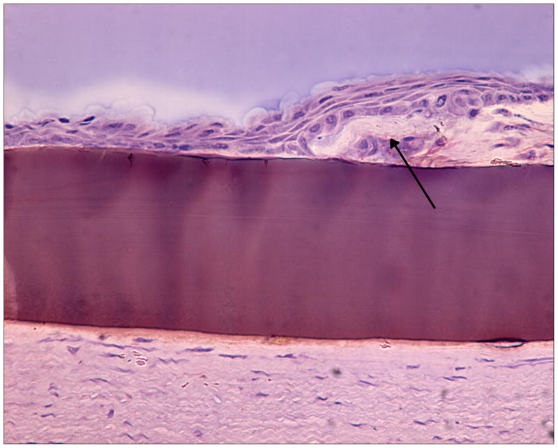
Figure 9a. Histological section showing epithelial overgrowth directly on a PEG/PAA hydrogel (central purple region, 100 microns thick) in the central part of a rabbit corneal onlay after 14 days. The arrow indicates the “ledge” of anterior cornea from which the epithelial cells are migrating.
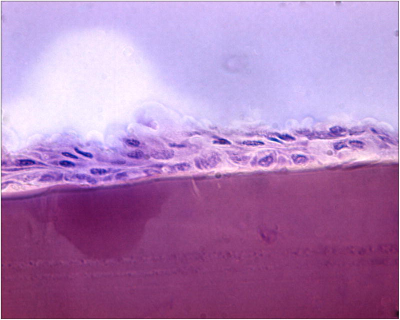
Figure 9b. A more central, magnified view of the histological section shown in Figure 9a.


