
Figure 4.
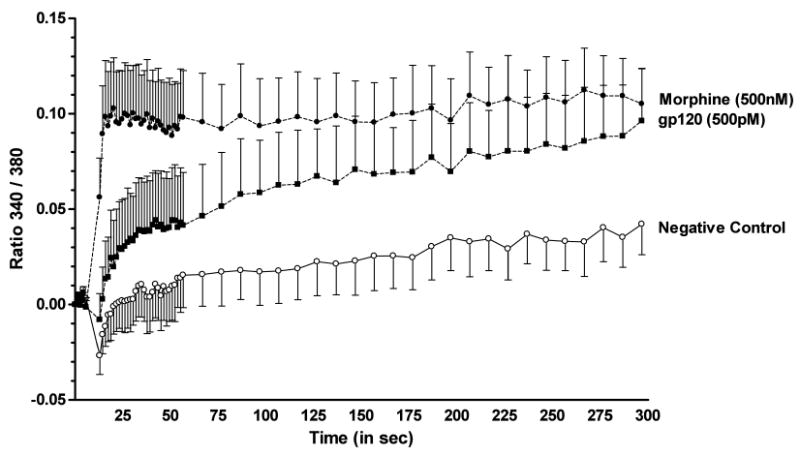
Analysis of [Ca2+]i activation in astrocytes after exposure to morphine or gp120. Cultures of enriched primary astrocytes derived from cerebral cortex were grown to 80-90% confluence, then loaded with 10μM fura-2/AM. Ratiometric Ca2+ measurements were made before (7.3 sec, 5 timepoints) and after (∼5 min) injection of morphine (500 nM, final concentration; filled circles) or gp120 (500 pM, final concentration; filled squares). Results are reported as the mean 340/380 ratio, and represent N = 6 samples per treatment group ± S.E.M. A two-way mixed ANOVA [treatment and time] showed that either morphine or gp120 significantly increased (p ≤ 0.001 and p ≤ 0.05, respectively) the 340/380 ratio compared to cells treated with vehicle (open circles). Morphine effects were noted immediately after exposure and remained stable over the 5 min recording period, while gp120 effects reached significance after 2 min.