
Fig. 2.
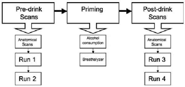
Schematic of the fMRI protocol. During the fMRI session, subjects underwent 2 sets of scanning. The first set of scans (“predrink scans”) were without alcohol priming and consisted of anatomical scans and a counterbalanced order of 2 fMRI runs. These scans were followed by a break from scanning (“priming”), where subjects were taken out of the magnet and allowed to stretch while consuming a priming dose of alcohol within 10 minutes followed by another 10 minutes to allow absorption. Subjects were then breathalyzed and their BACs recorded. They were immediately placed back into the scanner for the second set of scans (“postdrink scans”), which was identical to the first set.