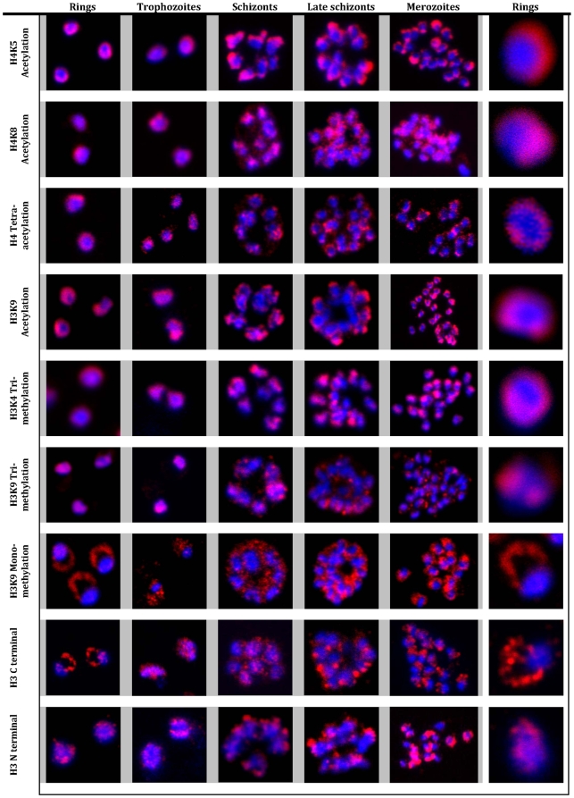
Figure 1. Immunofluorescence analysis of histone modifications in P. falciparum.
Localization of histone modifications were analyzed with the ring, trophozoite, schizont, late schizont and merozoite stages of the IDC. IFAs were carried out with antibodies against specific histone 3 and 4 lysine residue acetylations: H3K9Ac, H4K5Ac, H4K8Ac, and H4Ac4, as well as methylations: H3K4Me3, H3K9Me3 and H3K9Me1, and unmodified histone H3. Nuclear DNA was stained with DAPI (blue). All modifications, with the exception of H3K9Me1 and H3 (antibody raised against H3 C terminal), showed specific and distinct localization in the nucleus in all the stages. In contrast, H3K9Me1 was localized mainly outside the nucleus with very low levels detected inside the nucleus.

