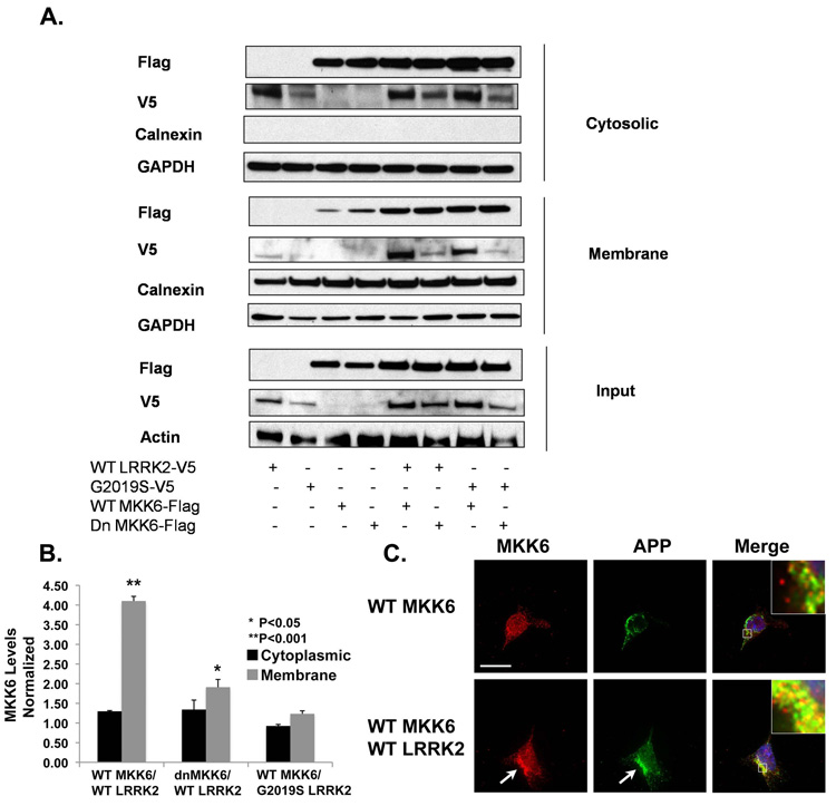
Fig. 4.
Expressing LRRK2 increases the level of MKK6 in the membrane fraction. (A) Both WT and G2019S LRRK2 increased the level of membrane MKK6. (B) Quantification indicated that the level of membrane MKK6 increased about 4-fold when expressed along with LRRK2 (*p<0.05; N=3, compared to WT MKK6). There was no significant difference in the level of membrane MKK6 between the WT or G2019S LRRK2-transfected samples. The level of MKK6 in the membrane fraction was normalized to the level of MKK6 in the total lysate, and compared between samples transfected ± LRRK2. Experiments were repeated three times in HEK-293FT cells. (C) Co-expression of WT and G2019S LRRK2 with WT MKK6 increased levels of MKK6 present at the plasma membrane, as shown by co-localization with amyloid precursor protein (APP). The arrows point to LRRK2/APP co-localization. Bar, 15 µm. All experiments have been performed three times.

