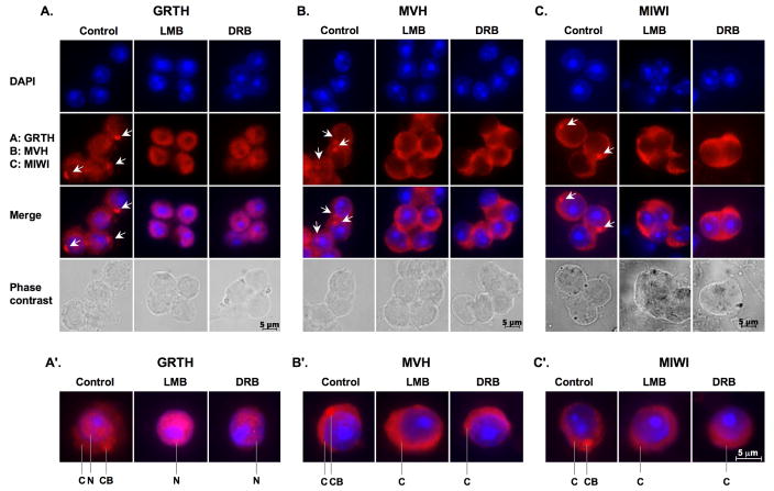
Fig. 2. Disappearance of GRTH and MVH and MIWI signal from CBs via a different mechanism after LMB and DRB treatment.
Spermatids isolated from stages VII–VIII were incubated with vehicle (control), nuclear protein export inhibitor (LMB) or RNA polymerase II inhibitor (DRB) for 3 h. Dried-down slides were immuno-stained with GRTH (A), or MVH (B) or MIWI antibodies (C) (2nd panel), nuclear staining by DAPI (1st panel), and merged image (3rd panel) and phase contrast (4th panel). Alexa Fluor 568 anti-IgG was used as a secondary antibody. Arrow-heads indicate the CBs. A′–C′: Single cell magnification of merged images. C: cytoplasm., N: Nucleus., CB: chromatoid body.

