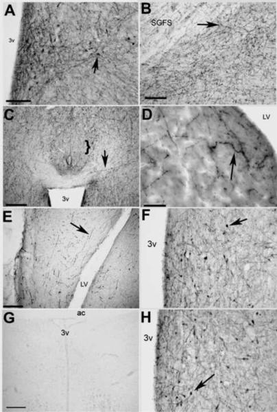
Fig. 2.
Representative photomicrographs of KP-lir. A) KP-lir cell bodies were only observed in the POM (bar = 30 μm). B) KP-lir fibers were observed in the optic tectum (bar = 30 μm). C) Dense KP-lir fibers were observed throughout the diencephalon, however, few--if any--fibers were observed in the bed nucleus of the pallial commisure (nCPa, bracket, bar = 50 μm.). D) KP-lir fibers were also observed in the septal area (bar = 20 μm), and E) in the hippocampus (bar = 50 μm). F) Preadsorption of sections with an unrelated peptide, galanin-like peptide, had no effect on KP-lir. G) Preadsorption of sections with excess kisspeptin eliminated KP-lir staining in the POM. Bar = 50 μm. H) Preadsorption of sections with a related RF-amide peptide, gonadotropin inhibitory peptide, also had no effect on KP-lir. Bar = 50 μm. 3v = third ventricle, LV = lateral ventricle, SGFS = striatum griseum et fibrosum superficiale, arrows designate positive KP-lir.

