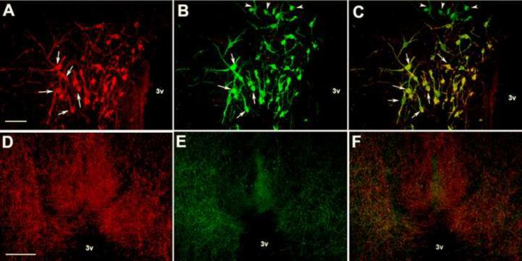
Fig. 4.
Representative photomicrographs of fluorescent immunocytochemistry for KP-lir (A, arrows), aromatase-ir (B, arrows). C) An overlay of kisspeptin- and aromatse-ir indicating colocalization of the two peptides (arrows). Bar = 50 μm. Representative photomicrographs of KP-lir fibers (D), GnRH-immunoreactive fibers (E) in the tuberoinfundibular region. F illustrates an overlay of panels D & E. Bar = 100 μm, 3v = third ventricle.

