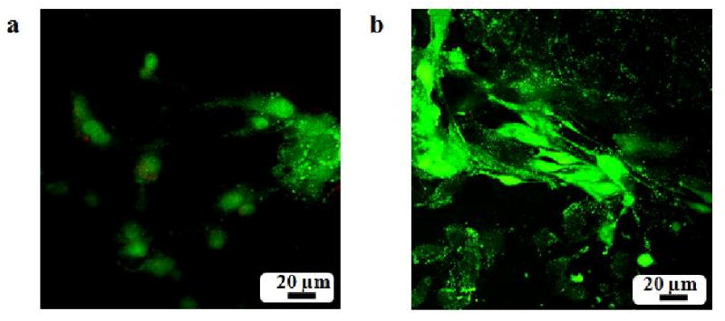
Figure 4.

Fluorescence images of PRO cells on polymer matrices with the Live/Dead cell stain after 48 hours of cell seeding. (a) PLAGA; (b) Matrix1. The osteoblast cells exhibit robust growth with a well-spread morphology on the blend surface compared to PLAGA.
