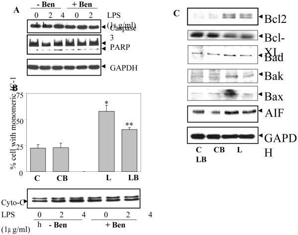
Fig. 3. Benfotiamine prevents LPS-induced activation of caspase-3, mitochondrial membrane potential (MMP) and Bcl-2 family proteins in RAW cells.
A. Growth-arrested RAW cells without or with Benfotiamine were incubated with 1 μg/ml of LPS for 24 h and Caspase-3 activation and PARP cleavage were determined in the cell lysate by western blot analysis using specific antibodies. A representative blot is shown (n=4). B. For assay of MMP, the growth-arrested RAW cells were treated with LPS (2.5 μg/mL) for 4h with or without benfotiamine. The cells were harvested and washed with PBS and MMP was evaluated by staining with JC-1 dye and analyzed with flow cytometry. Twenty thousand events were acquired for each sample. The data are means ± SD; (n=3). *p<0.0007 Vs Control; **p< 0.006 Vs LPS. Cytochrome-C release in the cytosol was measured after 2 and 4 h of LPS treatment by western blot using specific antibodies. GAPDH was used as loading control. C. The expression of Bcl-2 family proteins and AIF in the cell lysate from (A) was determined by western blot analysis using specific antibodies. A representative blot is shown (n=4). C, control; CB, control+benfotiamine; L, LPS; LB, LPS+benfotiamine.

