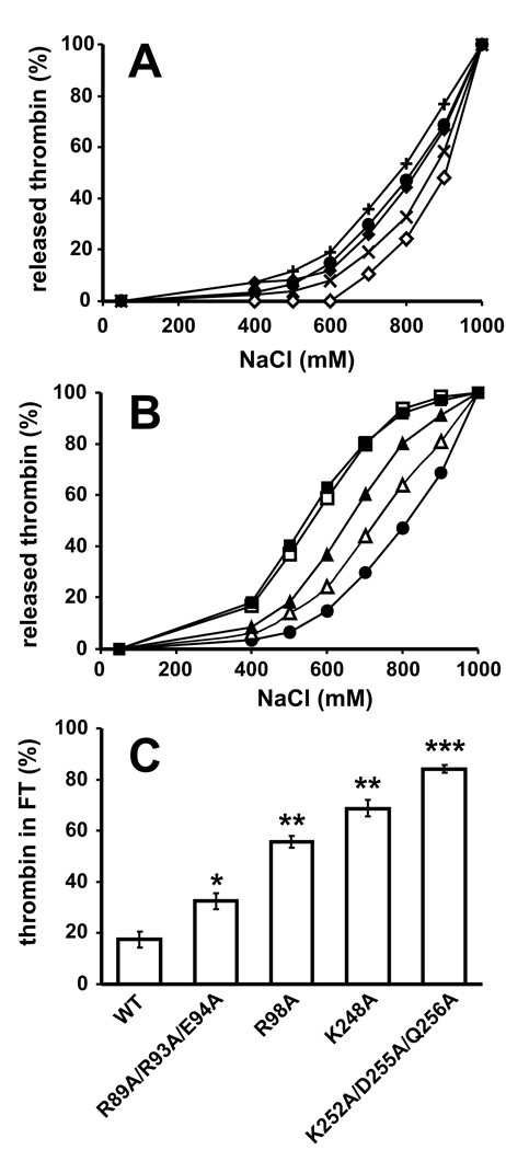
Fig. 4.
Exosite II mutants of thrombin show reduced binding to immobilized polyP. (A) 27 pmol WT (●), H66A (◆), Y71A (◇), E229A (×), or W50A (+) thrombins were incubated with polyP-zirconia beads, and bound enzyme was sequentially eluted with increasing NaCl concentrations (plotted as cumulative thrombin recovery). (B) 27 pmol WT (●), R89A/R93A/E94A (△), R98A (▲), K248A (□), or K252A/D255A/Q256A (■) thrombins were incubated and eluted as in panel A. (C) 27 pmol nM WT, R89A/R93A/E94A, R98A, K248A, or K252A/D255A/Q256A thrombins were incubated with polyP-zirconia beads in binding buffer containing 500 mM NaCl, with unbound thrombin in the flow-through expressed as percent of the starting thrombin concentration. The data shown in (A) and (B) are representative graphs of four separate experiments performed in duplicate and the values shown in (C) represent mean ± S.D. (n = 3). *P< 0.05, **P< 0.005, ***P< 0.0005.

