
Figure 9.
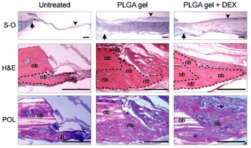
Photomicrographs of tissue sections prepared from untreated, PLGA colloidal gel (PLGA gel)-treated, or PLGA colloidal gel with encapsulated DEX (PLGA gel + DEX)-treated rat cranial bone defects. Top row: Low magnification of tissue sections including the bone defect margins (arrows) and the mid-portion of the defects (arrow heads). S-O, safranin-O and fast-green staining. Middle row: Higher magnification of tissue sections showing both the original host bone (ob) and the new bone (nb) formed in the areas adjacent to the host bone. New bone is outlined by a dotted line. H&E, hematoxylin and eosin staining. Bottom row: Polarizing photomicrographs (POL) of the cranial defect margins demonstrate that collagen fibers of the new bone (nb) display non- or low-polarizing orientation compared to that of the highly polarized parallel lamellae of the original host bone (ob) in each treatment group. H&E staining. Scale bar = 100 μm for all photos in this figure.