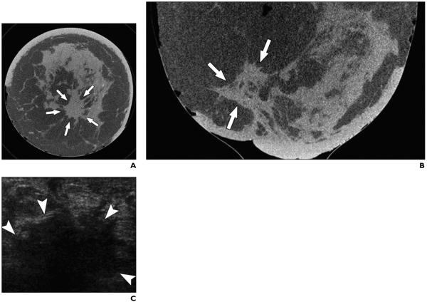
Fig. 3.
51-year-old woman with invasive ductal carcinoma of left breast occupying area of 6 × 5 cm.
A and B, Coronal (A) and axial (B) CT scans of left breast show irregular mass with spiculated margins (arrows). C, Transverse sonogram shows irregular solid hypoechoic mass in left retroareolar position with angular margins and dense posterior acoustic shadowing (arrowheads).

