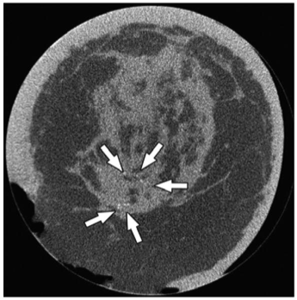
Fig. 4.

63-year-old woman with invasive ductal carcinoma of left breast. Coronal CT image shows microcalcifications within area of architectural distortion representing known cancer (arrows). Pathology showed ductal carcinoma in situ associated with microcalcifications.
