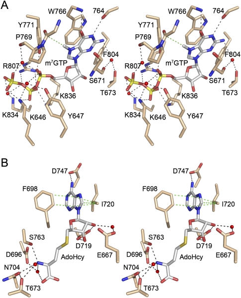
FIGURE 1.
Methyl acceptor and donor sites of human Tgs1. The figure shows a stereo view of the active site of human Tgs1 in complex with m7GTP (A) and AdoHcy (B) (from PDB 3GDH). (A) Highlighted are the hTgs1 side chains that contact the cap methyl acceptor, either directly, or via waters (depicted as red spheres). Hydrogen bonds and electrostatic interactions are denoted by black dashed lines. A single van der Waals contact between a main chain Cα and the guanine-N7 methyl group is depicted as a dashed green line. The m7G base is sandwiched between Trp766 and Ser671. (B) Highlighted are the hTgs1 side chains that contact the methyl donor. The adenine base of AdoHcy is sandwiched between Phe698 and Ile720, which make van der Waals contacts denoted by the dashed green lines.

