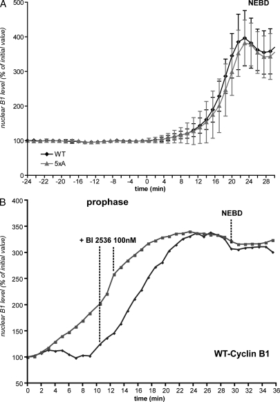
Figure 4.
Nuclear accumulation of cyclin B1 in prophase is independent of its phosphorylation and of Plk1 activity. (A) Cells coexpressing wild-type (WT) cyclin B1–mCherry and 5xA (Ser116, 126,128,133, and 147A)–cyclin B1–GFP were assayed, and the nuclear accumulation of the proteins was quantified. Mean curves of quantifications in different cells are displayed (n = 5). (B) Cells expressing wild-type cyclin B1–mCherry were assayed, and100 nM Plk1 inhibitor (BI 2536) was added during the nuclear import of cyclin B1 in prophase. Two examples are displayed (one image/minute). Error bars show SEM.

