Figure 5.
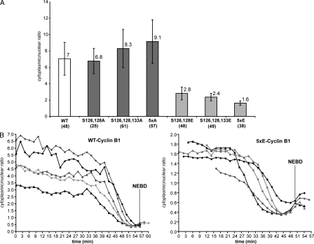
The nuclear/cytoplasmic distribution of cyclin B1 is only affected by phosphorylation in its N terminus domain in interphase. (A) The nuclear/cytoplasmic ratio of wild-type (WT) cyclin B1–GFP, Ser126– and 128A–cyclin B1–GFP, Ser126–,128A–, and 133A–cyclin B1–GFP, 5xA (Ser116,126,128,133, and 147A)–cyclin B1–GFP, Ser126– and 128E–cyclin B1–GFP, Ser126–,128A–, and 133E–cyclin B1–GFP, and 5xE (Ser116,126,128,133, and 147E) cyclin B1–GFP was quantified on optical sections of asynchronous interphase HeLa cells. Mean values ± SEM and numbers of cells assayed are displayed. (B) Cells expressing either wild-type or 5xE cyclin B1–GFP were recorded. Real time changes in the nuclear/cytoplasmic ratio were quantified on optical sections as cells entered mitosis. Five examples are displayed for each experiment.

