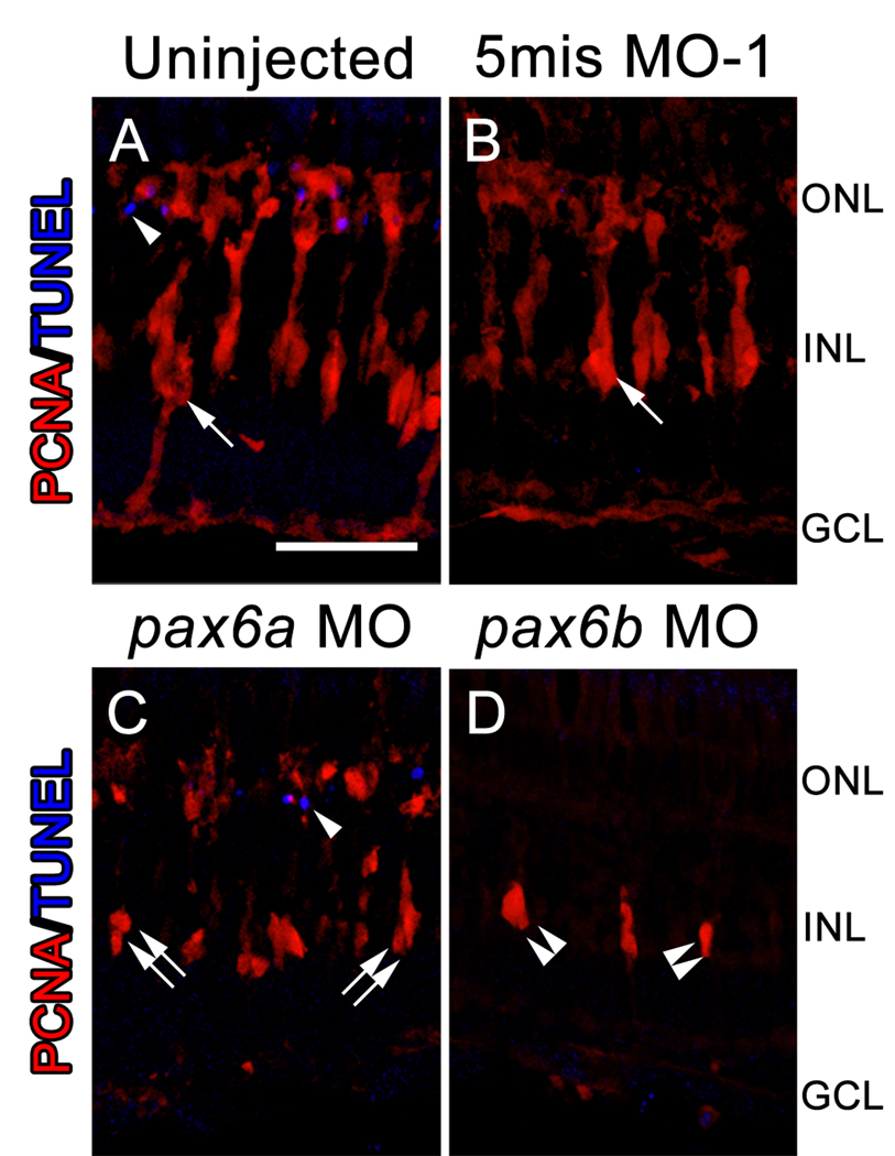
Figure 5. Reduced numbers of INL neuronal progenitors observed in the pax6 morphants is not a result of increased cell death.
Dark-adapted adult albino zebrafish were either uninjected or injected and electroporated with a 5-base mismatch control morpholino (5mis MO-1), anti-pax6a morpholino, or anti-pax6b morpholino. PCNA expression (red) and TUNEL labeling (blue) were assessed after 72 hours of constant light treatment by immunohistochemistry. The uninjected and 5mis MO-1 retinas (A, B) both contained large columns of PCNA-positive neuronal progenitor cells (arrow). However, knockdown of only Pax6a (C) yielded columns (double arrows) that contained fewer PCNA-positive cells relative to controls. In contrast, knockdown of only Pax6b (D) produced clusters of neuronal progenitors (double arrowheads) that were fewer in number than even the pax6a morphant retina. TUNEL-positive nuclei (single arrowheads) were occasionally observed in the ONL of both the control and pax6 morphant retinas (A, C). However, no TUNEL-positive nuclei were observed in the INL of either the control or pax6 morphant retinas. The scale bar in panel A represents 25 microns and is the same for panels B–D. GCL, ganglion cell layer; INL, inner nuclear cell layer; ONL, outer nuclear layer.

