
Figure 2. Developmental role of abd-A expression in the P compartment .
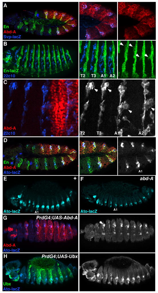
A. Lateral view of a stage 11 svp-lacZ Drosophila embryo immunostained for Abd-A (red), En (green), and β-gal (blue). svp-lacZ expression serves as an early marker for oenocytes. B. Stage 16 en-lacZ Drosophila embryo immunostained for β-gal (green) and with a PNS-specific neuronal marker (mAb22C10, blue). Arrows (in right panel) point to where the dch3 (in T2/T3) and lch5 (A1/A2) sensory organs form within the P compartment. C. Close up view of the T2 and T3 thoracic and A1 and A2 abdominal segments immuostained with Abd-A (red) and mAb22C10 (blue, black and white at right). D. Lateral view of a stage 11 ato-lacZ embryo immunostained for Abd-A (red), En (green) and β-gal (blue, black and white at right). Note that the majority of ato-lacZ expression in the thorax and abdomen is within the P compartment. E. Wild type ato-lacZ embryo (stage 14) immunostained for β-gal (blue). Note the higher levels of ato-lacZ within the abdomen than the thorax. F. ato-lacZ expression is the same in all body segments of abd-A− embryos. G. PrdG4;UAS-Abd-A embryos show that Abd-A (red) expression within the thorax stimulates ato-lacZ activity (blue, black and white at right). H. PrdG4;UAS-Ubx embryos show that ectopic Ubx (green) does not alter ato-lacZ levels within the thorax.