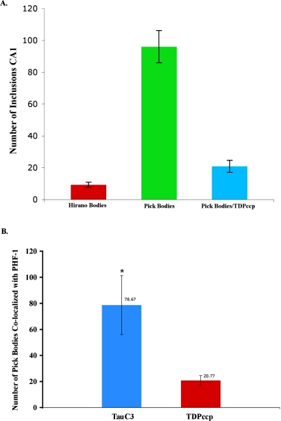
Figure 3.
Quantification of Pick bodies double-labeled by TDPccp, TauC3, and PHF-1. In both A and B, PHF-1 labeling was utilized as a marker for Pick bodies. A: Data show the average number of Hirano bodies labeled with TDPccp, Pick bodies labeled with PHF-1, and Pick bodies with both PHF-1 and TDPccp, identified in a 20X field of area CA1 (n=3 fields for 3 different Pick disease cases) ±S.E.M. B: a semi-quantitative analysis was performed to estimate the total number of PHF-1-labeled Pick bodies that co-localized with either TDPccp or TauC3 within the CA1 region of the hippocampus (percent given above bars). Results indicated the total number of double-labeled Pick bodies with caspase-cleaved tau plus phosphorylation was significantly greater compared with caspase-cleaved TDP-43 plus phosphorylation. Data represent the average (±S.E.M.) of three different fields from three representative Pick cases (χ2 = 3.857, p-value* < x 0.05).

